Clinical practice · 7 min read
Recognising arrhythmias on the ECG: three questions
Narrow or wide QRS, regular or irregular, and how P waves relate to QRS: three questions that sort arrhythmias on ECG into AV blocks, atrial fibrillation and flutter, SVT and ventricular tachycardia.
- Published
In this article
- Arrhythmias on ECG: patient, rate, then three questions
- Slow rhythms: the P–QRS relation names the AV block
- Narrow-complex tachycardia: atrial fibrillation, flutter or SVT
- Wide QRS: think ventricular tachycardia first
- Common pitfalls
- Worked example: three questions on one strip
- Why mix ECGs when you practise
- How to practise on MedIQ
Night shift: the monitor alarms, showing a fast rhythm at about 150/min. No time for long lists; you need short questions, asked in the same order every time. However complex arrhythmias on ECG strips look, three questions sort out most of them: narrow or wide QRS, regular or irregular rhythm, and the P–QRS relation.
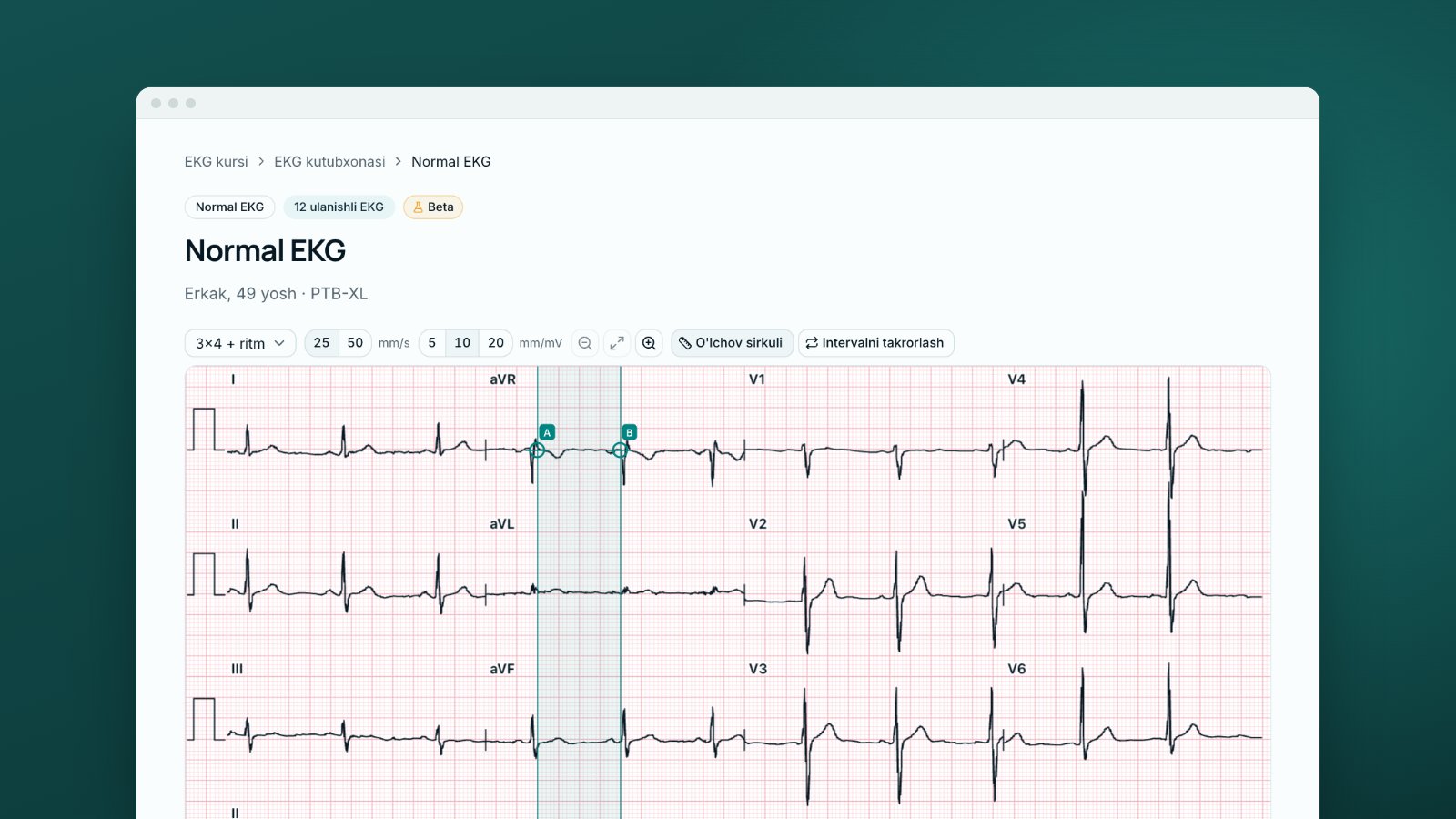
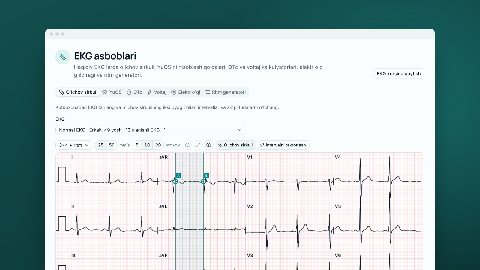
This post covers the questions, main rhythms, pitfalls and what mixed practice offers. Rate and intervals are covered in ECG measurements, the overall order in reading an ECG systematically.
Arrhythmias on ECG: patient, rate, then three questions
Check the patient first: an unstable patient's treatment does not wait for the ECG analysis (see below). Then take the rate: tachycardia is above 100/min [1]. The bradycardia cut-off varies: 60/min in many definitions, 50/min in the 2018 ACC/AHA/HRS guideline [2].
- Narrow or wide QRS? A narrow QRS (≤120 ms) usually indicates normal ventricular activation through the His–Purkinje system; a rare exception is idiopathic ventricular tachycardia [1]. Causes of wide-QRS (>120 ms) tachycardia are in the table [1].
- Regular or irregular? Compare RR intervals with calipers across the whole strip, not just two cycles.
- How do P waves relate to the QRS? Is there a P before each QRS, is the PR constant, are there extra P waves? In a tachycardia, an atrial rate above the ventricular rate usually means flutter or atrial tachycardia [1]; in a slow rhythm it may point to AV block (below).
The first two questions sort tachycardias into four groups [1]:
| QRS | Regular | Irregular |
|---|---|---|
| Narrow | Sinus tachycardia, atrial tachycardia, flutter with fixed AV conduction, AV nodal re-entrant tachycardia (AVNRT), orthodromic atrioventricular re-entrant tachycardia (AVRT) | Atrial fibrillation, multifocal atrial tachycardia, flutter with variable conduction |
| Wide | Ventricular tachycardia; supraventricular tachycardia (SVT) with bundle branch block; conduction over an accessory pathway; ventricular pacing; SVT with QRS widened by electrolyte disturbance or antiarrhythmic drugs | Atrial fibrillation, or flutter with variable conduction, with bundle branch block or aberrancy; pre-excited atrial fibrillation; polymorphic ventricular tachycardia |
Slow rhythms: the P–QRS relation names the AV block
The 2018 ACC/AHA/HRS definitions of second-degree AV block require P waves at a constant rate below 100/min [2]:
| Finding | P–QRS relation [2] |
|---|---|
| First-degree AV block | Every P conducts, PR >200 ms; a delay rather than a block |
| Second-degree AV block, Mobitz type I (Wenckebach) | PR lengthens until one P is not conducted; grouped QRS complexes |
| Second-degree AV block, Mobitz type II | Constant PR before and after the non-conducted P |
| Second-degree AV block with 2:1 conduction | Every other P conducts; cannot be classed as Mobitz I or II |
| High-grade AV block | Two or more consecutive P waves blocked, some conducted |
| Third-degree (complete) AV block | No AV conduction; usually a junctional or ventricular escape rhythm |
The level of block sets the risk. Nodal block progresses more slowly and has a more reliable, atropine-responsive escape rhythm. Block within or below the His bundle can progress rapidly and unexpectedly, with a slower escape rhythm that does not respond to atropine [2]. Mobitz I with a narrow QRS is most likely nodal [2].
Narrow-complex tachycardia: atrial fibrillation, flutter or SVT
Atrial fibrillation. No discernible regular P waves; irregular ventricular activation with no RR pattern unless AV block is present [4]. The 2024 ESC guideline requires ECG documentation: a 12-lead ECG, or at least 30 seconds on a single- or multi-lead device (a consensus threshold) [4].
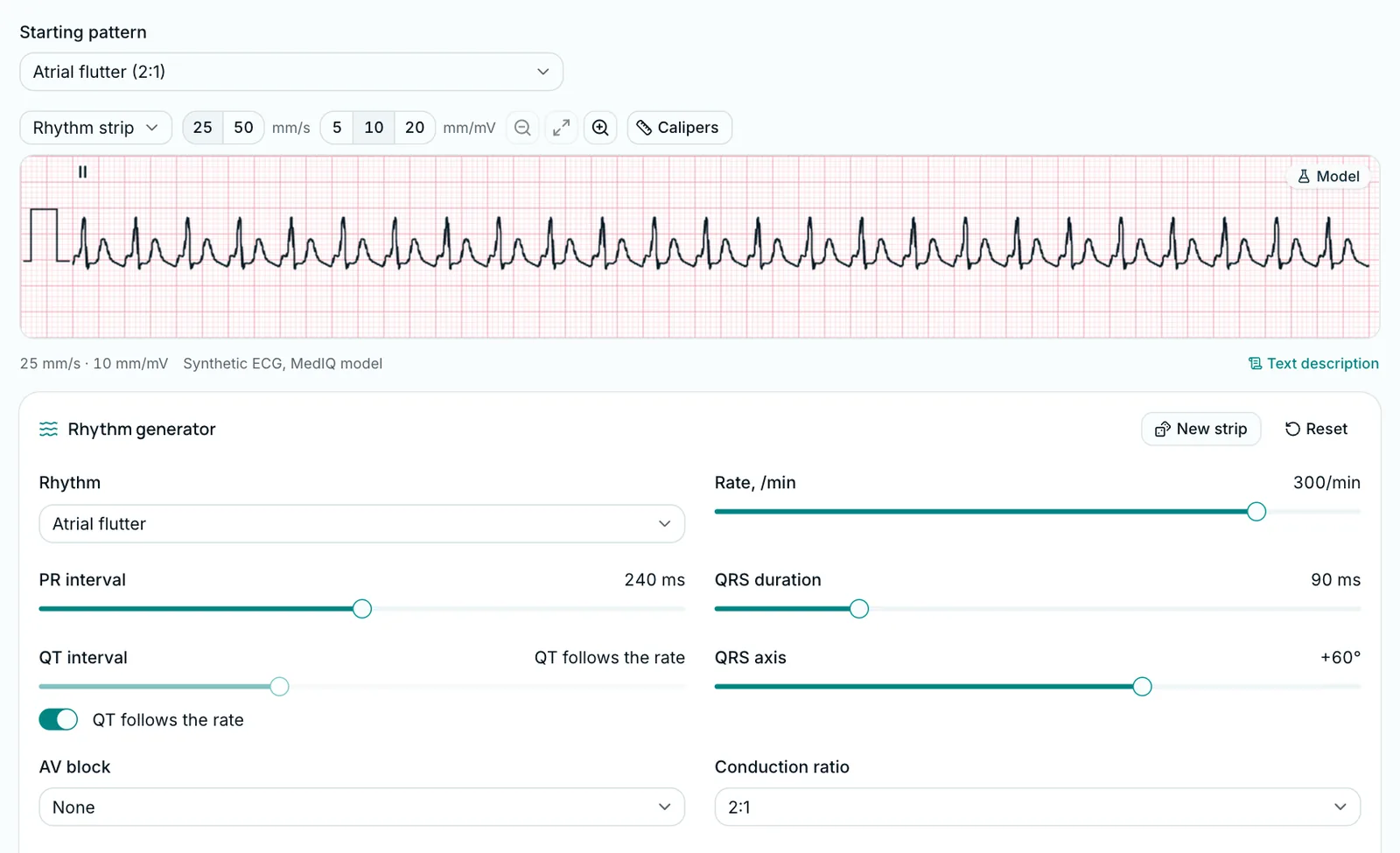
Atrial flutter. Regular atrial activation at 250–330/min; in typical (counter-clockwise) flutter, saw-tooth F waves are negative in II, III and aVF and positive in V1 [1]. A regular narrow-complex tachycardia at about 150/min suggests 2:1 flutter [1].
AVNRT and AVRT. In typical AVNRT the retrograde P hides in the QRS or appears as a small wave at its end: pseudo-r′ in V1, pseudo-S inferiorly. In orthodromic AVRT the RP interval is constant. Vagal manoeuvres and adenosine may aid the diagnosis [1].
Wide QRS: think ventricular tachycardia first
The 2022 ESC guideline defines ventricular tachycardia (VT) as three or more consecutive ventricular beats above 100/min [5]. It is non-sustained up to 30 seconds, sustained if it lasts at least 30 seconds or needs intervention to stop. Monomorphic VT keeps one QRS shape; polymorphic VT changes continually [5].
The rule: a wide-QRS tachycardia is VT until proven otherwise [1]. AV dissociation, capture beats and fusion beats are its key features [1]. An irregular wide-QRS tachycardia suggests atrial fibrillation with bundle branch block or aberrancy, pre-excited atrial fibrillation, or polymorphic VT [1].
The patient comes first. In the 2022 ESC guideline, sustained monomorphic VT with haemodynamic instability needs immediate synchronised cardioversion, or an unsynchronised shock if synchronisation is not possible [5]. Without a pulse it is cardiac arrest: pulseless VT is defibrillated by the resuscitation algorithm, as for ventricular fibrillation [7]. Record a haemodynamically tolerated tachycardia on a 12-lead ECG [5].
Common pitfalls
- Fast atrial fibrillation looks regular. At high rates the irregularity is hard to see and can pass for a regular SVT [1].
- A regular rhythm at about 150/min. With 2:1 conduction, flutter may not be obvious on the ECG [1].
- A blocked premature atrial complex. In atrial bigeminy, non-conducted premature beats can be misread as AV block [2]. March out P–P with calipers.
- Classifying a 2:1 block. With only 2:1 conduction, it cannot be called Mobitz I or II [2].
- Atrial fibrillation with a slow, regular ventricular rhythm. A regular rate below 50/min suggests complete AV block [2].
Worked example: three questions on one strip
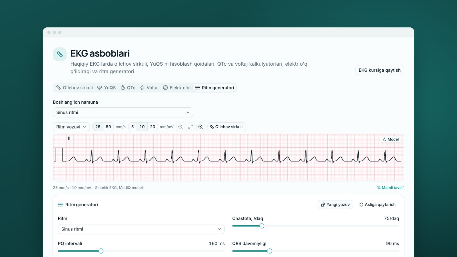
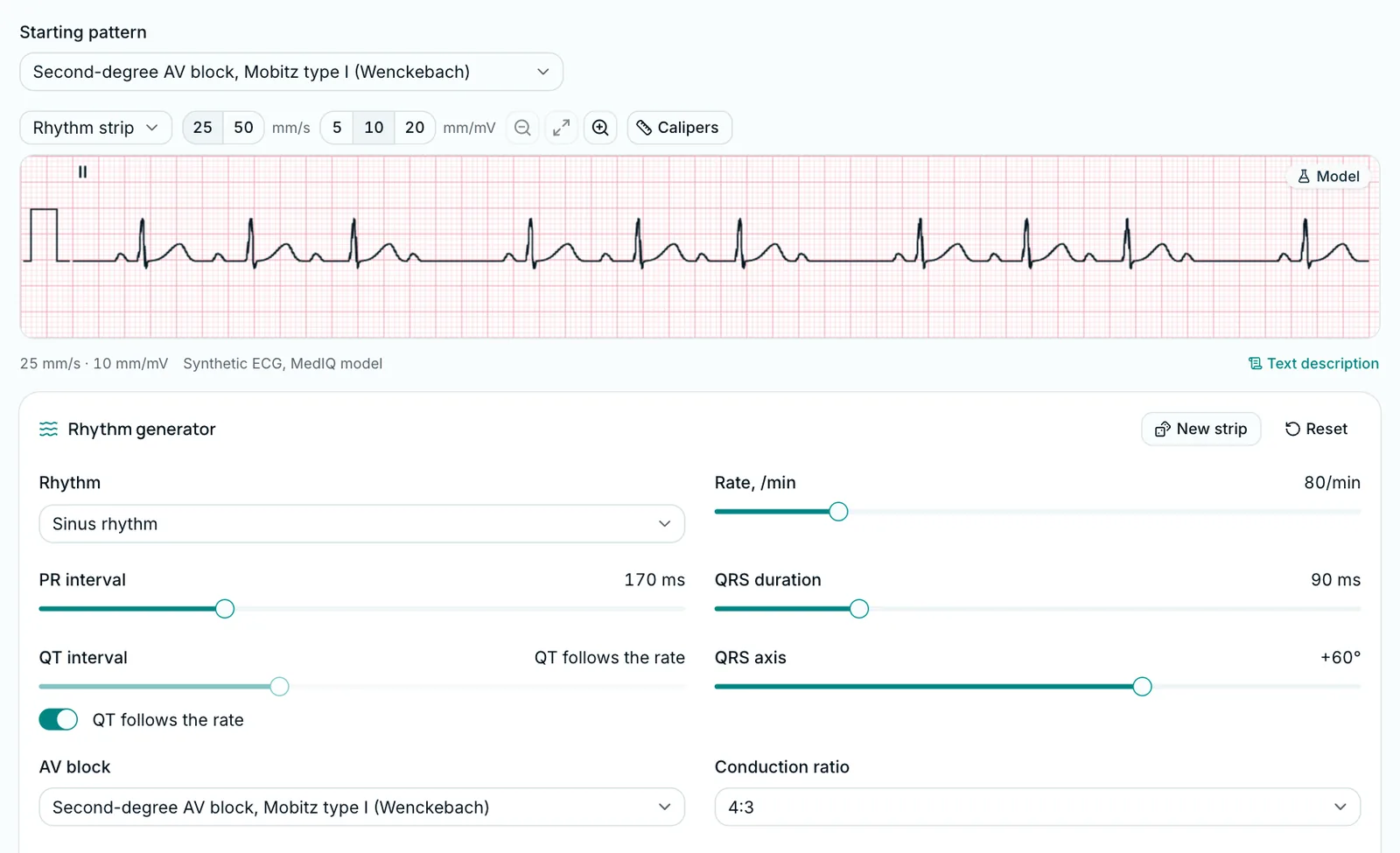
Open the "Second-degree AV block, Mobitz type I (Wenckebach)" pattern in the Rhythm generator: a Model strip in lead II.
- Rate: P waves at a steady 80/min, ventricles at 60/min on average.
- QRS: narrow.
- Rhythm: irregular but not chaotic: three QRS complexes, a pause, three more.
- P and QRS: constant P–P, three QRS per four P waves. The PR lengthens each cycle (here 170 to about 300 ms) until the fourth P is blocked; after the pause it is short again.
Conclusion: "Sinus rhythm, second-degree AV block, Mobitz type I, 4:3 conduction". Switch the AV block to Mobitz II: the PR stays constant, single P waves suddenly drop (in practice Mobitz II more often comes with a wide QRS [2]). Then choose 2:1: can the strip alone give the type? No [2].
Why mix ECGs when you practise
In Hatala and colleagues' second experiment, 66 first-year students had the same instruction and then practised in two groups: one saw different diagnoses mixed, the other each diagnosis in its own block. On new ECGs, accuracy was 46% after mixed and 30% after blocked practice [3]. It was a single two-hour session, the groups were not strictly randomised, and the diagnoses were ventricular hypertrophy, bundle branch blocks, myocardial infarction, ischaemia and pericarditis, not rhythms [3]. In a 2022 meta-analysis, the pooled result of studies of this contrastive approach was not statistically significant [8]. For rhythms, mixing is a sensible principle rather than a proven rule.
In a block you know the category in advance and only recognise the feature; mixed practice makes you decide the diagnosis each time, as at the monitor [3]. Our post on spaced repetition covers this too.
How to practise on MedIQ
The Rhythm generator (ECG tools page and many lessons, e.g. 26 and 29–34) has more than 50 model rhythms and patterns. Change the rate, AV block, conduction ratio or extrasystoles and press "New strip". Every strip carries the "Model" badge: model-generated, not recorded from a patient.
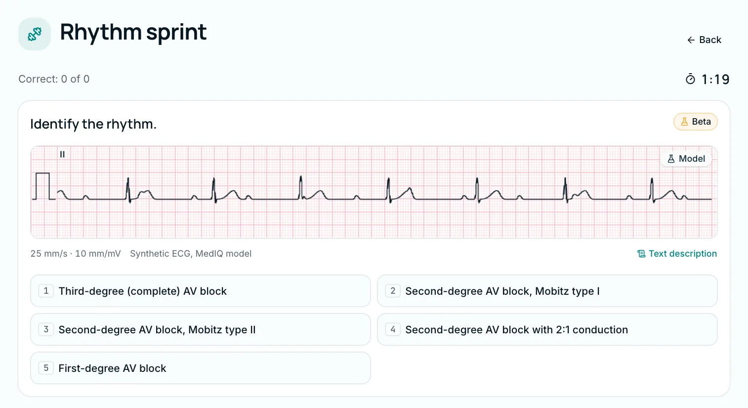
In the Rhythm sprint you name as many rhythms as you can in 90 seconds, one answer per strip, with the result shown at once. Rhythm types come mixed, not in blocks; strips you have not answered in the last 30 days come first. Most are Model strips; the rest are mainly real MIT-BIH recordings, each reviewed independently by two cardiologists [6]. The diagnosis shown is the dataset's own label.
Related lessons: "First- and second-degree AV block", "Complete (third-degree) heart block", "Atrial fibrillation", "Atrial flutter", "Narrow-complex tachycardias" and "Ventricular tachycardia". The ECG course is in Beta: checked by MedIQ AI, not yet by a physician. Found a mistake? Press "Report an error". More on the ECG course page.
This post is educational; real-patient decisions follow local protocols and the responsible physician.
Sources
- 1.Brugada J, Katritsis DG, Arbelo E, Arribas F, Bax JJ, Blomström-Lundqvist C, et al. 2019 ESC Guidelines for the management of patients with supraventricular tachycardia. Eur Heart J. 2020;41(5):655–720. doi.org/10.1093/eurheartj/ehz467
- 2.Kusumoto FM, Schoenfeld MH, Barrett C, Edgerton JR, Ellenbogen KA, Gold MR, et al. 2018 ACC/AHA/HRS guideline on the evaluation and management of patients with bradycardia and cardiac conduction delay. Circulation. 2019;140(8):e382–e482. doi.org/10.1161/CIR.0000000000000628
- 3.Hatala RM, Brooks LR, Norman GR. Practice makes perfect: the critical role of mixed practice in the acquisition of ECG interpretation skills. Adv Health Sci Educ Theory Pract. 2003;8(1):17–26. doi.org/10.1023/A:1022687404380
- 4.Van Gelder IC, Rienstra M, Bunting KV, Casado-Arroyo R, Caso V, Crijns HJGM, et al. 2024 ESC Guidelines for the management of atrial fibrillation developed in collaboration with the European Association for Cardio-Thoracic Surgery (EACTS). Eur Heart J. 2024;45(36):3314–3414. doi.org/10.1093/eurheartj/ehae176
- 5.Zeppenfeld K, Tfelt-Hansen J, de Riva M, Winkel BG, Behr ER, Blom NA, et al. 2022 ESC Guidelines for the management of patients with ventricular arrhythmias and the prevention of sudden cardiac death. Eur Heart J. 2022;43(40):3997–4126. doi.org/10.1093/eurheartj/ehac262
- 6.Moody GB, Mark RG. MIT-BIH Arrhythmia Database (version 1.0.0). PhysioNet. 2005. doi.org/10.13026/C2F305
- 7.Soar J, Yeung J, Couper K, Deakin C, DeBono J, Nolan J, et al. Adult advanced life support guidelines. Resuscitation Council UK; 2025. www.resus.org.uk/professional-library/2025-resuscitation-guidelines/adult-advanced-life-support-guidelines
- 8.Oh SY, Cook DA, Van Gerven PWM, Nicholson J, Fairbrother H, Smeenk FWJM, Pusic MV. Physician training for electrocardiogram interpretation: a systematic review and meta-analysis. Acad Med. 2022;97(4):593–602. doi.org/10.1097/ACM.0000000000004607
This article is written for educational purposes only and is not a basis for clinical decisions. When treating a patient, rely on current clinical guidelines and the judgement of the responsible physician.
Tags
- arrhythmias on ECG
- AV block
- atrial fibrillation
- ventricular tachycardia
- ECG interpretation
- rhythm sprint
About the author
MedIQ Editorial
Editorial team for medical education
MedIQ Editorial is the team of clinicians and medical-education specialists who prepare the platform's learning content. Articles are based on current clinical guidelines (WHO, FIGO, ACOG, RCOG, NICE, ESHRE) and peer-reviewed research; sources are listed at the end of every article.