ECG course · Beta

Learn to read an ECG with confidence

See in 3D where every wave comes from, measure on real ECGs and practise every day. Lessons in Uzbek, Russian and English.

The ECG course is part of PRO. Verify your account to try it free for 3 days.

interactive lessons
35
real ECGs
198
practice items
477

Course map

Three levels, from the basics to clinical ECG

The first release has the lessons students need most. The rest follow in later releases.

Level A

ECG fundamentals

17 lessons

  1. Cardiac electrophysiology: where the ECG comes from

    • 1The conduction system of the heart
    • 3From cell to skin: dipole, vector and lead

    2 more lessons in a later release

  2. Recording the ECG: leads, electrodes and paper

    • 5Limb leads and the hexaxial reference system
    • 6Precordial leads and correct electrode placement
    • 7Paper, calibration and filters
    • 8Artefacts and lead reversals
  3. The normal ECG: waves, intervals, rate and axis

    • 9Waves, segments and intervals
    • 10Heart rate
    • 11Sinus rhythm and its variants
    • 12The electrical axis
    • 13R-wave progression and normal Q waves

    1 more lesson in a later release

  4. Systematic reading and the report

    • 15The 10-step ECG reading
    • 16QT and QTc
    • 17Writing the conclusion
  5. Atrial enlargement and ventricular hypertrophy

    • 18Atrial abnormality: reading the P wave
    • 19Left ventricular hypertrophy: voltage criteria and their limits
    • 20Right ventricular hypertrophy and the ECG in lung disease
Level B

Clinical ECG

17 lessons

  1. Intraventricular conduction: bundle branch and fascicular blocks

    • 21Right bundle branch block
    • 22Left bundle branch block
    • 24Ventricular pre-excitation: the WPW pattern and syndrome

    1 more lesson in a later release

  2. Bradyarrhythmias and AV block

    • 26First- and second-degree AV block
    • 27Complete (third-degree) heart block

    2 more lessons in a later release

  3. Supraventricular arrhythmias

    • 29Premature atrial and junctional complexes, junctional rhythms and multifocal atrial tachycardia
    • 30Atrial fibrillation
    • 31Atrial flutter
    • 32Narrow-complex tachycardias
  4. Ventricular arrhythmias and wide-complex tachycardia

    • 33Premature ventricular complexes (PVCs)
    • 34Ventricular tachycardia
    • 36Polymorphic VT, torsades de pointes and ventricular fibrillation

    1 more lesson in a later release

  5. Ischaemia and myocardial infarction

    • 37Ischaemia and injury: what the ST segment and T wave tell you
    • 38STEMI criteria: the Fourth Universal Definition
    • 39Localising the infarct and the culprit artery
    • 43STEMI or mimic? Other causes of ST elevation

    3 more lessons in a later release

  6. Electrolytes, drugs and systemic conditions

    • 44Potassium and the ECG: hyperkalaemia and hypokalaemia

    5 more lessons in a later release

Level C

Advanced ECG

1 lesson

  1. Inherited arrhythmia syndromes and sudden cardiac death

    In a later release

  2. Pacemakers and implantable devices

    In a later release

  3. Monitoring, Holter and exercise testing

    In a later release

  4. Emergency ECG: cardiac arrest and peri-arrest rhythms

    • 59Cardiac arrest rhythms

    2 more lessons in a later release

  5. Special populations: children, pregnancy, older adults

    In a later release

Inside the course

Seven tools, one goal: reading the ECG yourself

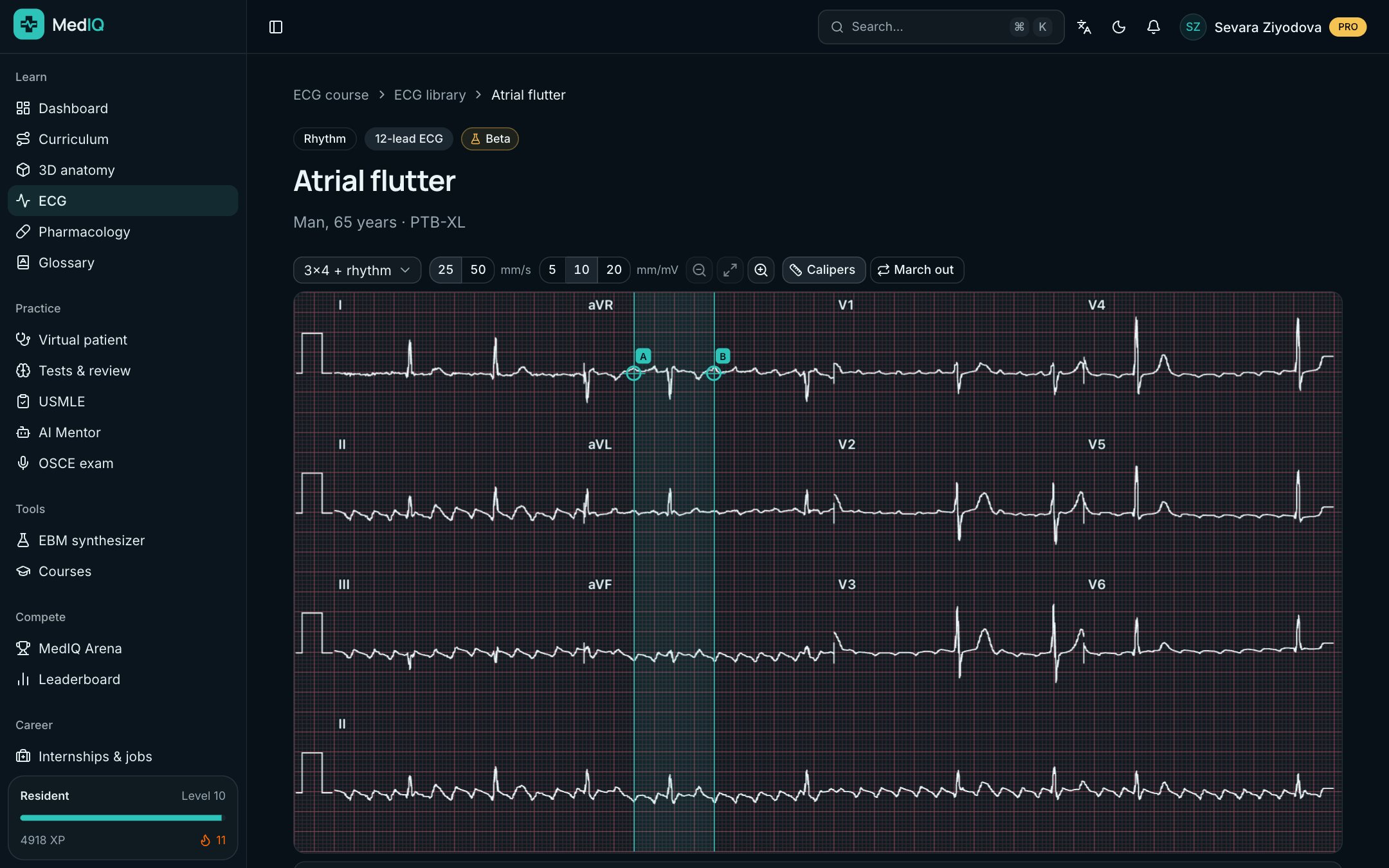
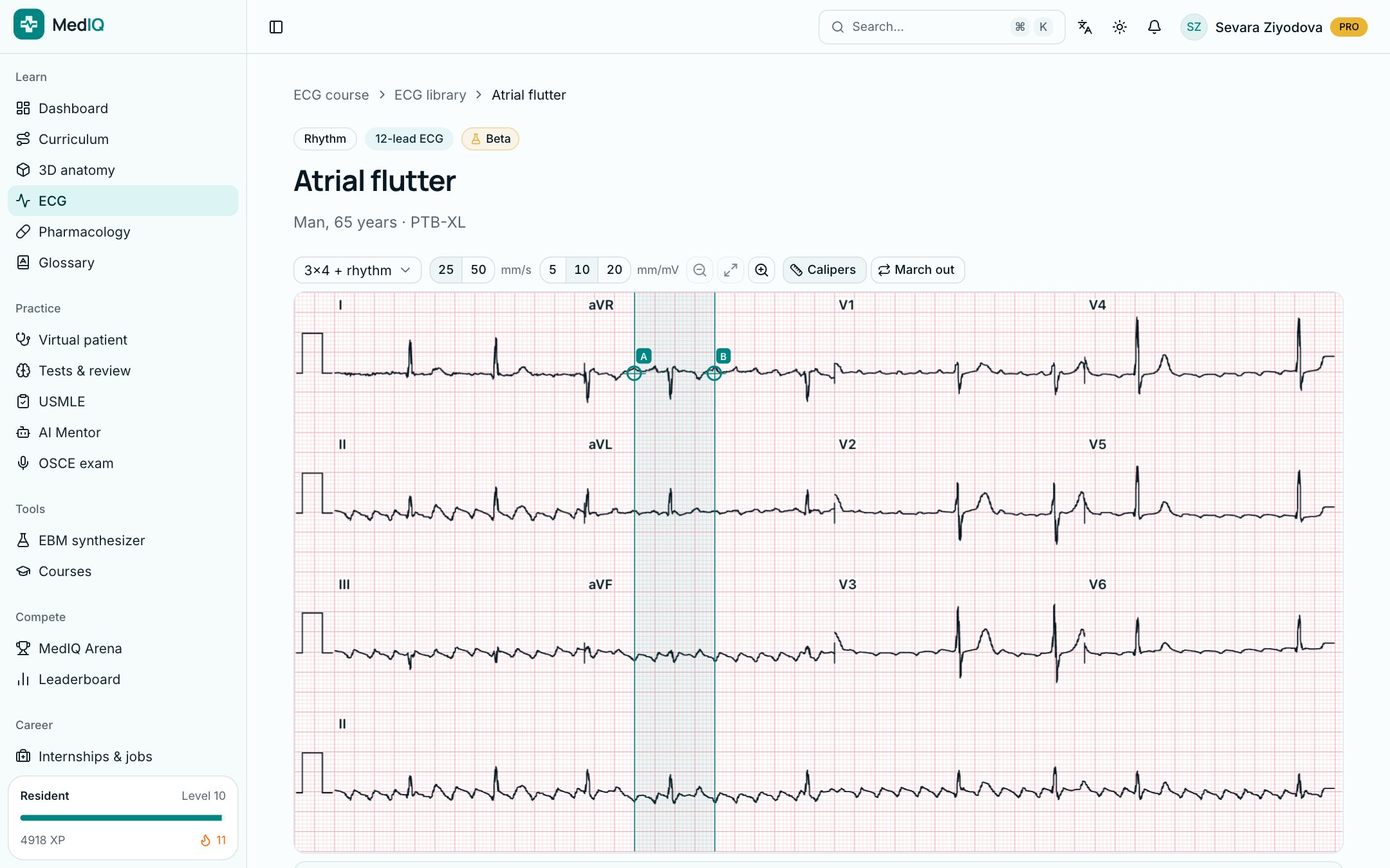
12-lead ECG

Read a real ECG as if on paper

Every lesson shows the ECG on millimetre grid paper with calibration and paper speed. Zoom into a lead, measure with calipers and compare your conclusion with the interpretation.

  • A systematic read: rhythm, rate, intervals, axis, waves
  • The Full read drill grades your report
  • Questions inside the lesson with instant feedback
12-lead ECG: a screen of the MedIQ ECG course
ECG: PTB-XL (CC BY 4.0)

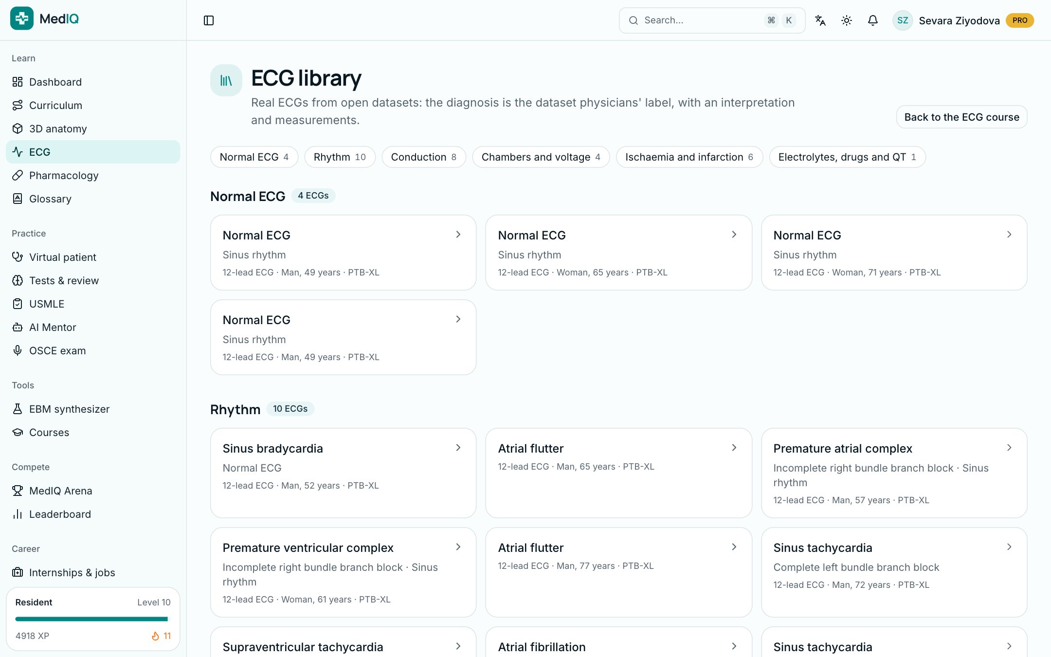
ECG library

Real recordings diagnosed by physicians

The library's ECGs come from open clinical datasets, sorted by topic. The diagnosis is the dataset physicians' label; MedIQ AI never changes it.

  • 12-lead ECGs and rhythm strips
  • A conclusion, measurements and an interpretation for each
  • Source and licence under every ECG
ECG library: a screen of the MedIQ ECG course

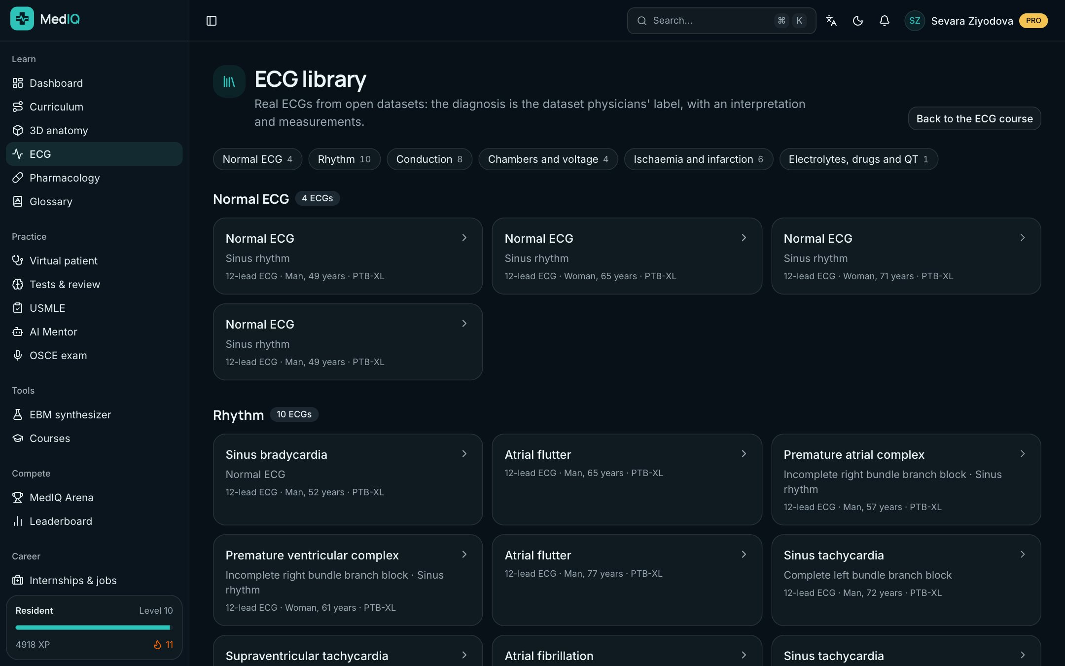
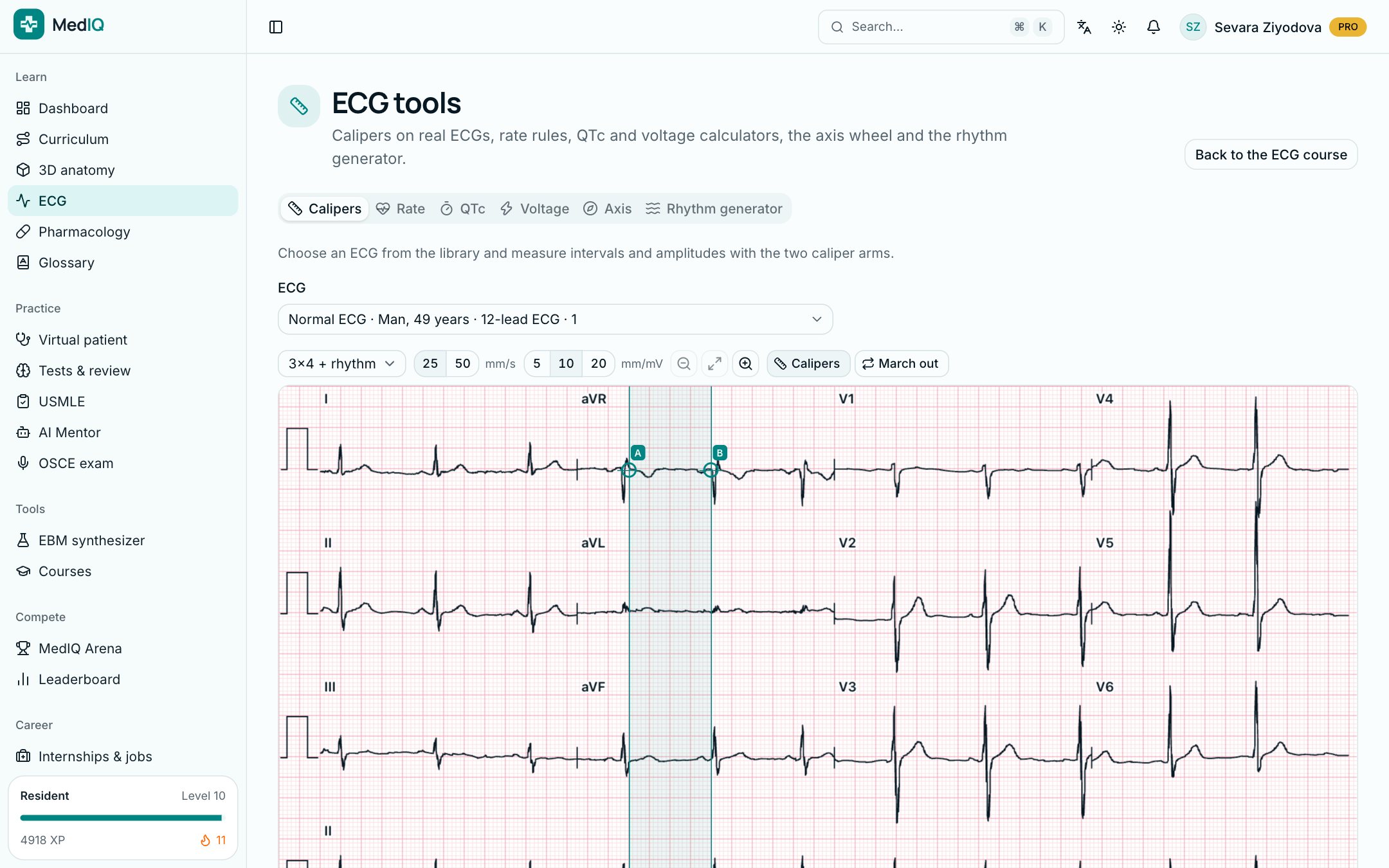
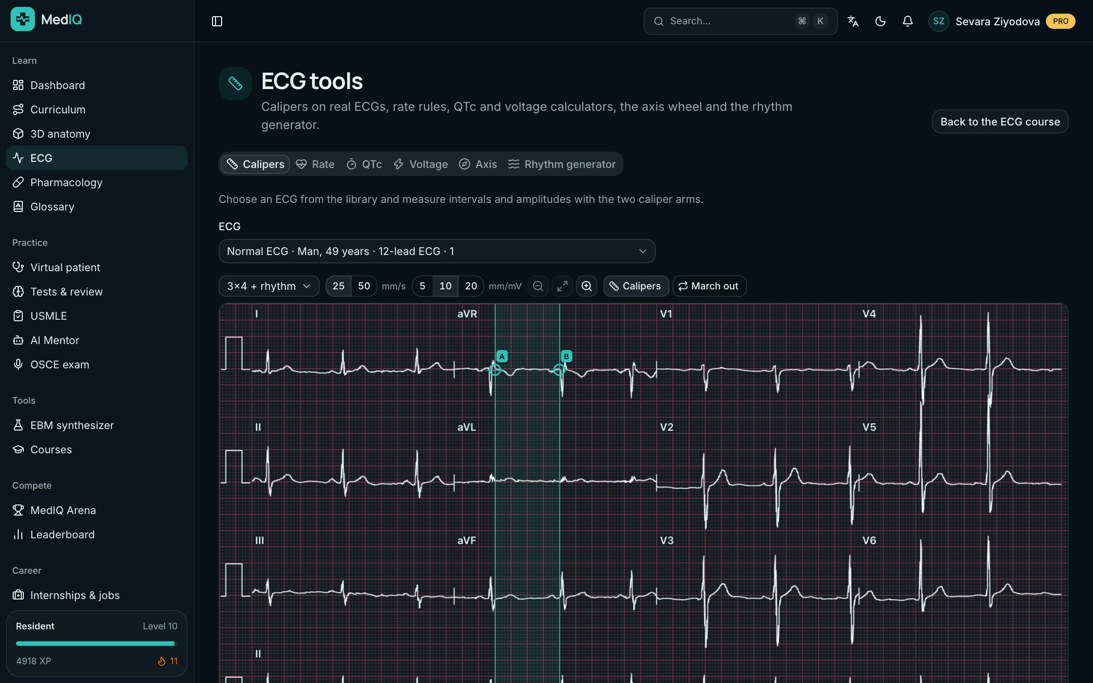
ECG tools

Measure and calculate

Measure intervals and amplitudes on a real ECG with calipers. The rate, QTc, voltage and axis calculators compare your result with normal values.

  • Calipers and a rate ruler
  • QTc with Bazett and Fridericia
  • A rhythm generator: build a rhythm yourself
ECG tools: a screen of the MedIQ ECG course
ECG: PTB-XL (CC BY 4.0)

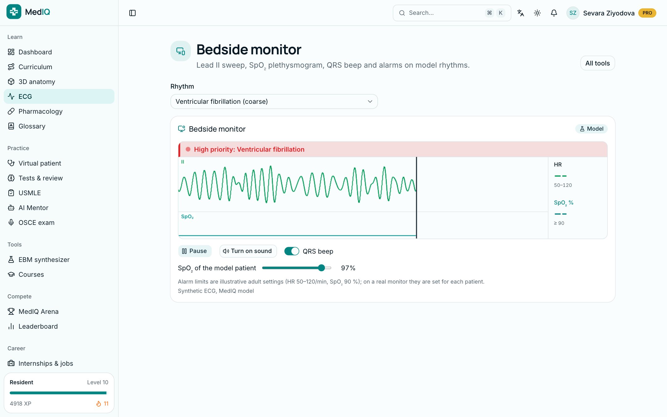
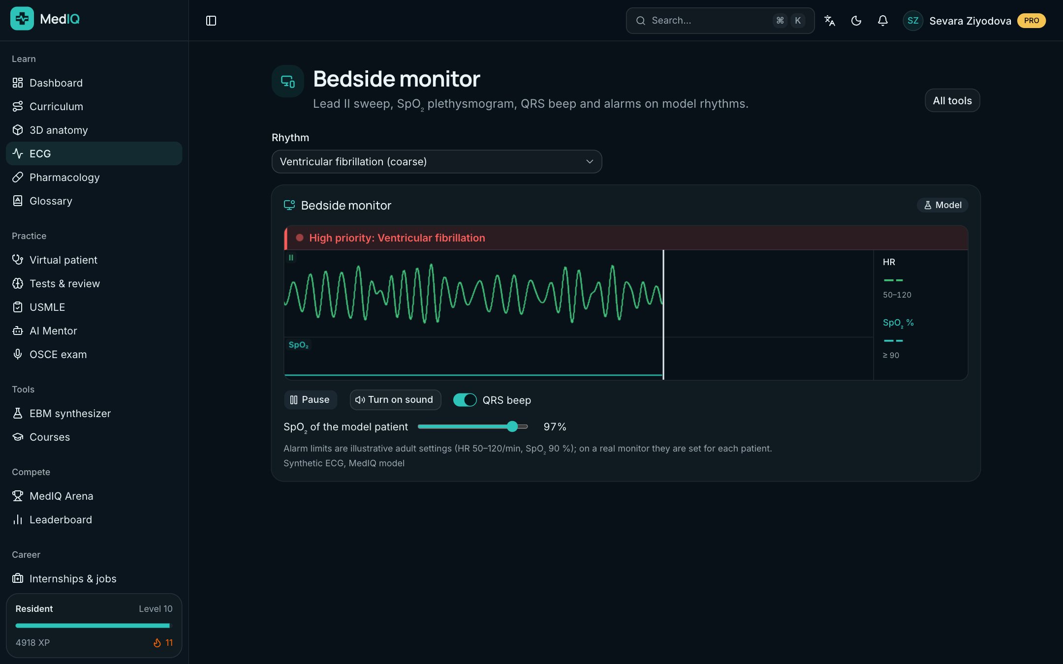
Bedside monitor

See and hear the rhythm

The monitor shows lead II, the SpO₂ pleth wave and a beep for every QRS. See how the alarms behave on model rhythms.

  • QRS beep and alarms
  • Cardiac arrest rhythms on a model patient
  • Learn to read a monitor screen
Bedside monitor: a screen of the MedIQ ECG course

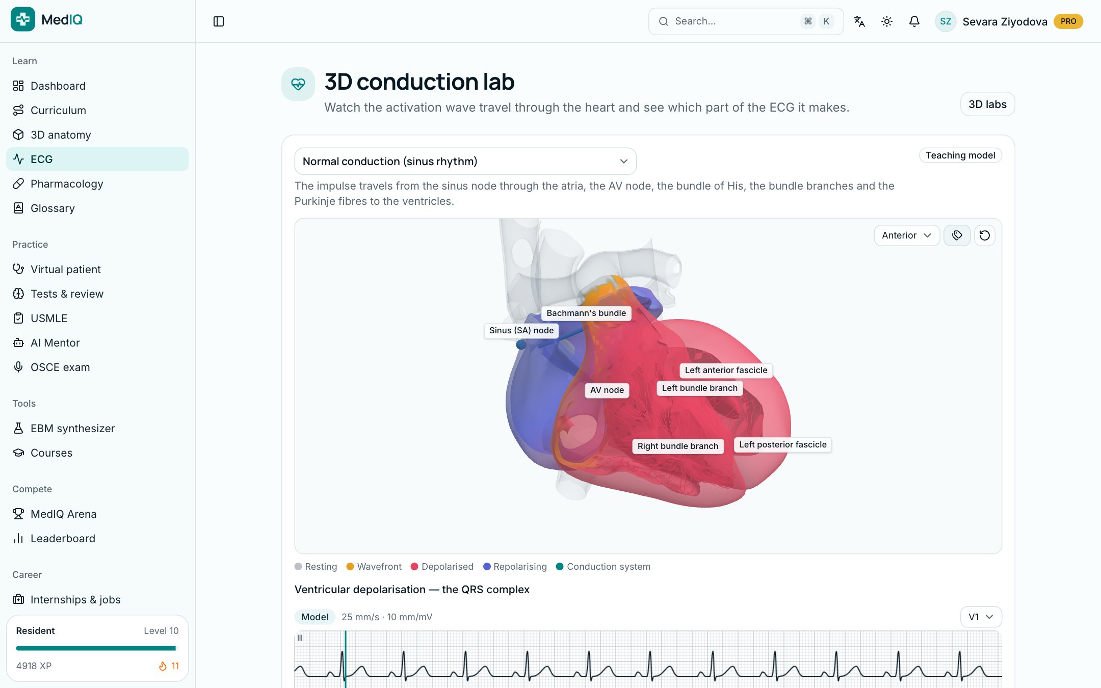
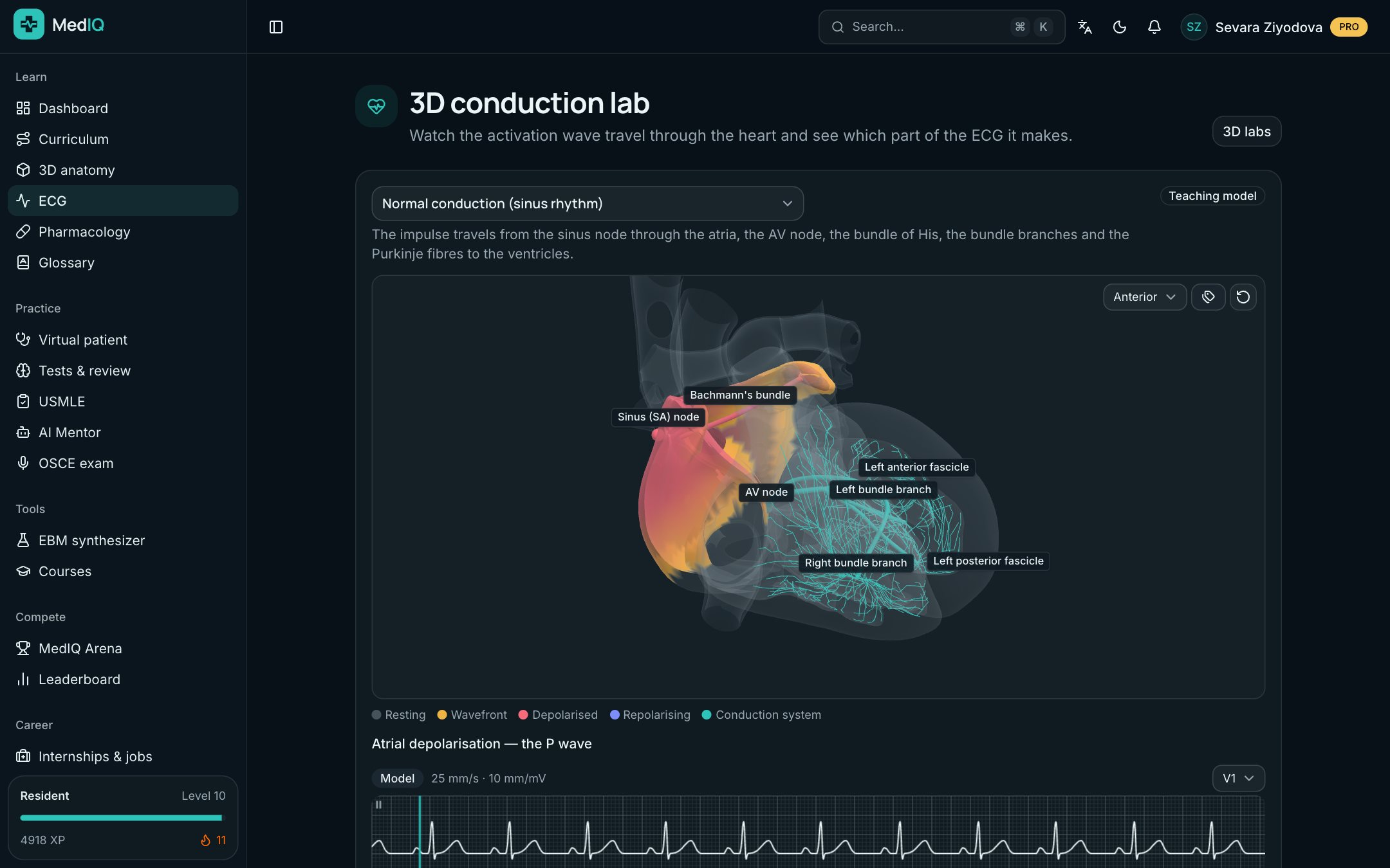
3D lab

Where the ECG waves come from

Watch in 3D how the wave of excitation travels from the sinus node to the Purkinje fibres, and which part of the ECG each step makes.

  • Normal rhythm, blocks, WPW, atrial flutter
  • The wave and the ECG trace side by side
  • Step-by-step playback and pause
3D lab: a screen of the MedIQ ECG course

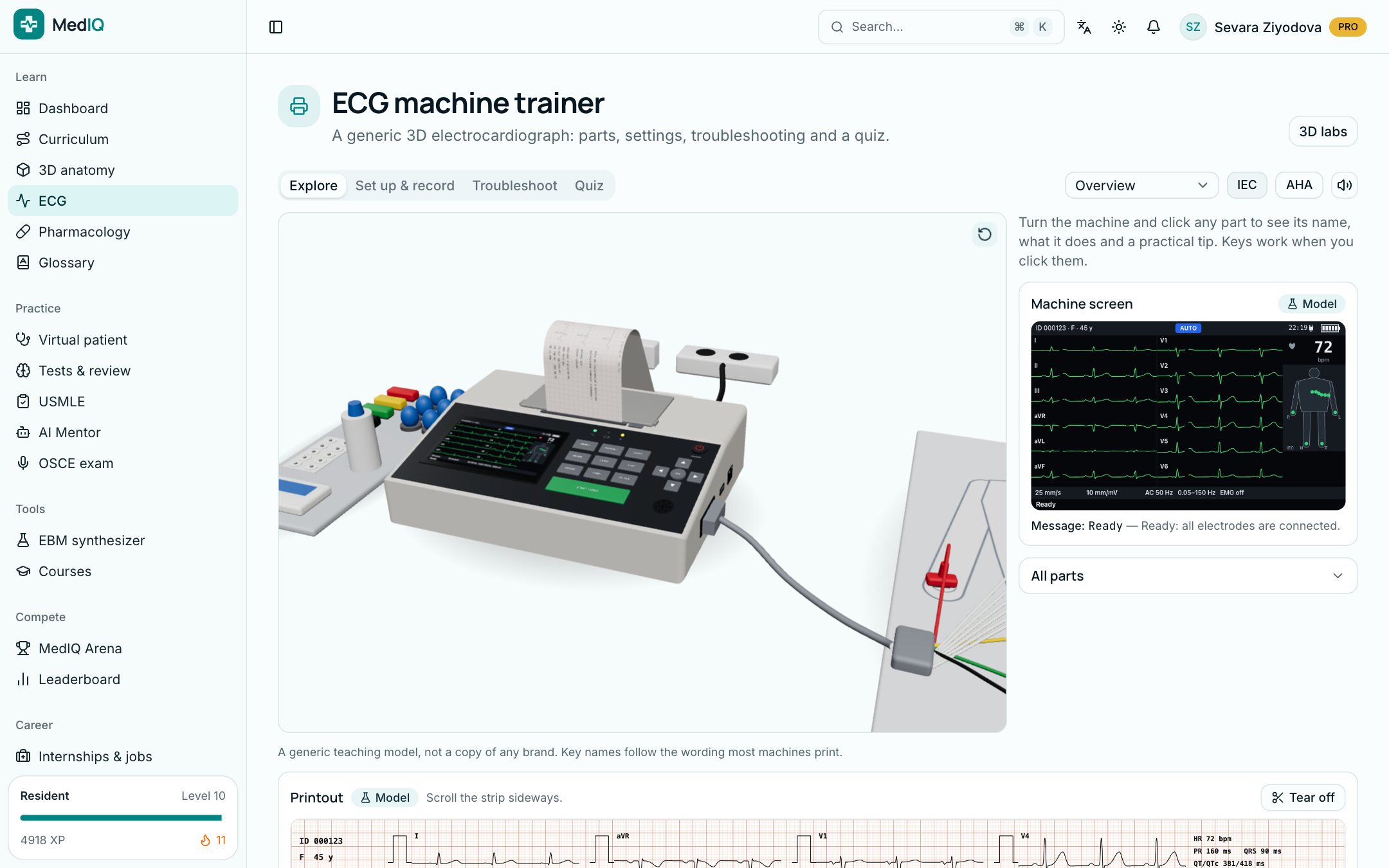
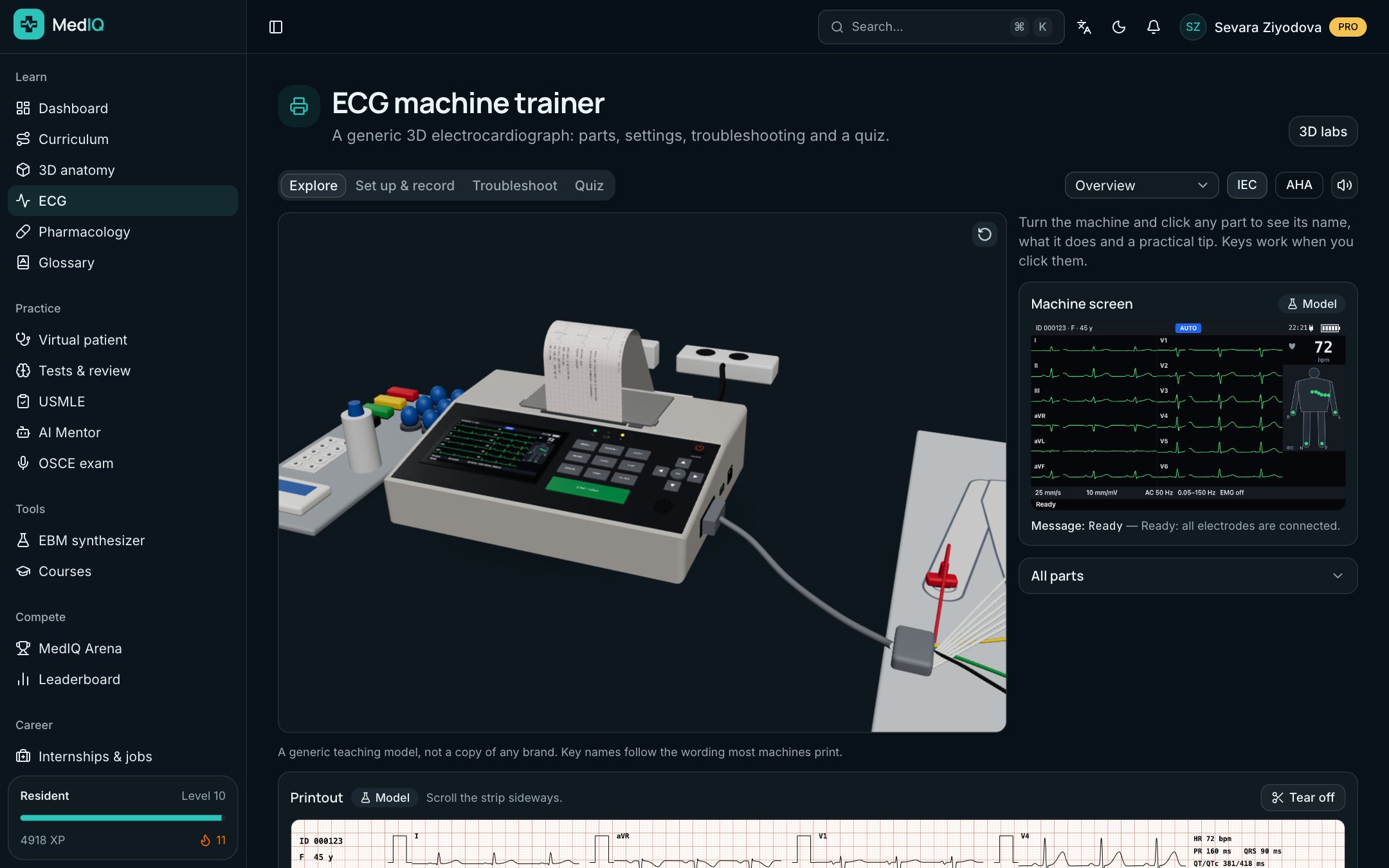
ECG machine trainer

Learn to record an ECG correctly

On a generic 3D electrocardiograph, learn the parts, set paper speed, gain and filters, and find and fix recording faults.

  • Electrodes and cable colours
  • Settings: speed, gain, filters
  • Troubleshooting and a quiz
ECG machine trainer: a screen of the MedIQ ECG course

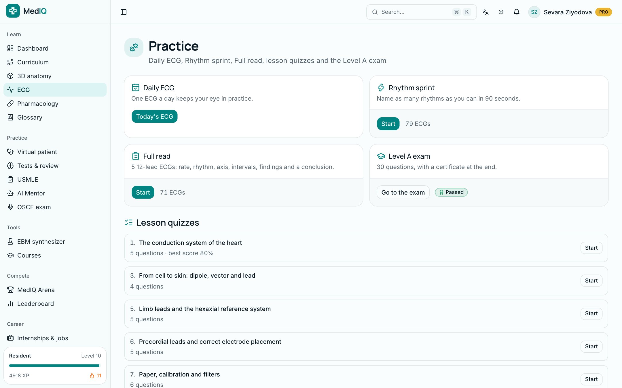
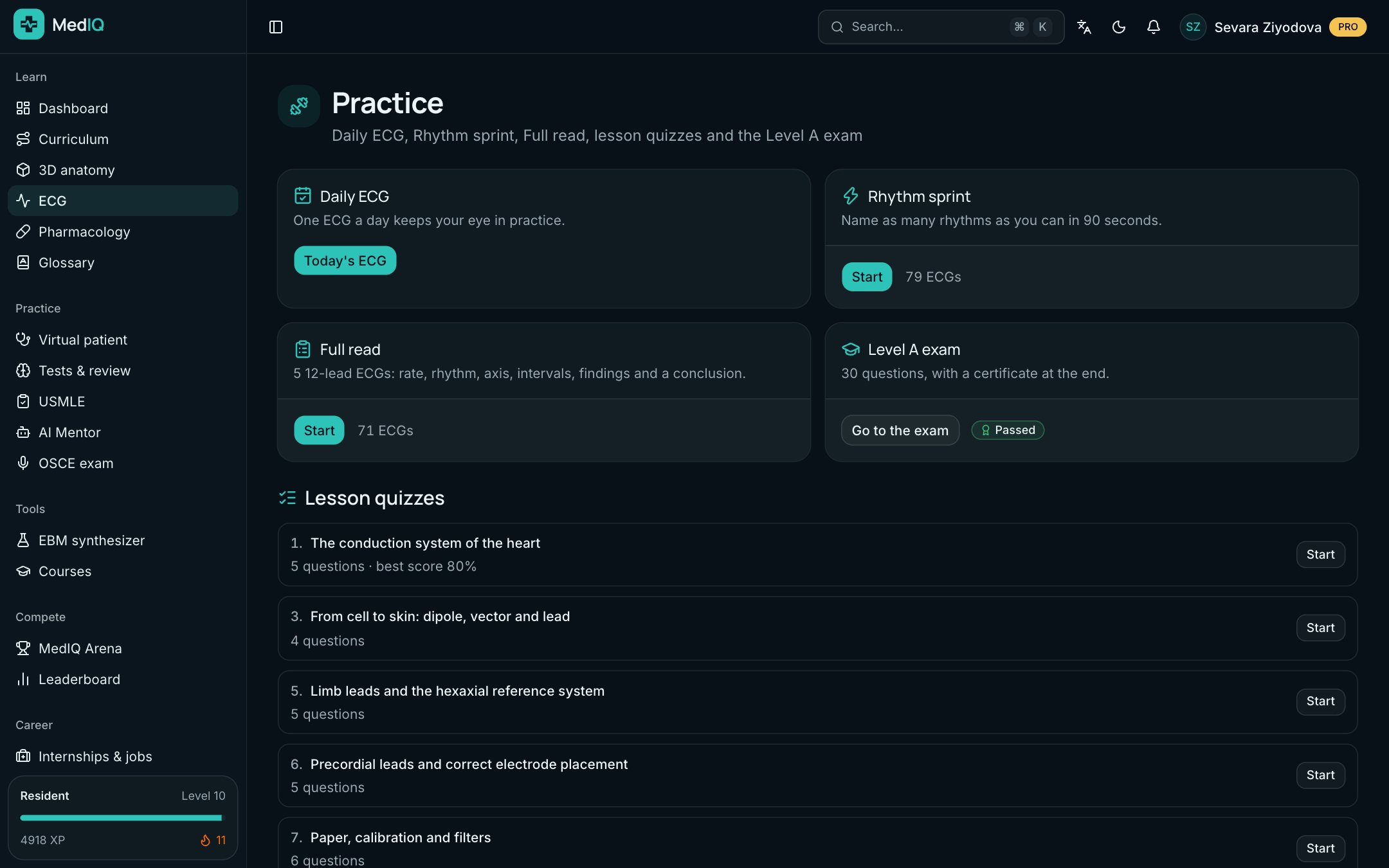
Practice and certificate

A little every day, with a certificate at the end

The daily ECG keeps your eye in, while Rhythm sprint and Full read build speed and accuracy. Pass the Level A exam to earn the "ECG fundamentals" certificate.

  • The daily ECG and your streak
  • Rhythm sprint and Full read drills
  • Level A exam: 30 questions, 45 minutes
Practice and certificate: a screen of the MedIQ ECG course

Quality and sources

Real data, open sources, honest labels

  • Open clinical datasets

    The real ECGs come from open datasets that allow commercial use; the source and licence are shown under every recording.

  • Diagnoses from physicians

    A real ECG's diagnosis is the dataset physicians' label. MedIQ AI never re-diagnoses it.

  • The "Model" label

    Synthetic recordings from the rhythm generator and the simulators are always labelled "Model": they are not a patient's ECG.

  • Beta and the report button

    MedIQ AI checked the content twice, independently; a physician has not reviewed it yet. Every page has a button to report an error.

ECG sources in the course

  • PTB-XLCC BY 4.0PTB-XL, Wagner et al.; PTB-XL+, Strodthoff et al.; PhysioNet, CC BY 4.0, modified
  • STAFF IIIODC-By 1.0STAFF III Database, Martínez et al., PhysioNet, ODC-By 1.0, modified
  • MIT-BIH ArrhythmiaODC-By 1.0MIT-BIH Arrhythmia Database, Moody & Mark, PhysioNet, ODC-By 1.0, modified
  • LUDBODC-By 1.0LUDB, Kalyakulina et al., PhysioNet, ODC-By 1.0, modified
  • CU Ventricular TachyarrhythmiaODC-By 1.0CU Ventricular Tachyarrhythmia Database, Nolle et al., PhysioNet, ODC-By 1.0, modified

Beta: the course content was checked by MedIQ AI and has not yet been reviewed by a physician. If you spot an error, report it with the button on the page.

Questions about the ECG course

Medical students, residents and young doctors. Level A starts from the ECG fundamentals, so no preparation is needed.

See what MedIQ can do for you.Today.